A
inflamação aguda é uma rápida resposta do hospedeiro que serve para levar
leucócitos e proteínas plasmáticas,tais como anticorpos,para os locais de
infecção ou tecido injuriado.A inflamação aguda tem 3 principais componentes:
(1)alterações no calibre vascular que levam a um aumento do fluxo sanguíneo;(2)
mudanças estruturais na microvasculatura que permitam a saída das proteínas
plasmáticas e os leucócitos da circulação(predominantemente polimorfos
nucleares) e (3) emigração de leucócitos
da microcirculação,seu acúmulo no foco da injúria e sua ativação para eliminar
o agente agressor.
O
apêndice é um divertículo verdadeiro normal do ceco que está sujeito a
inflamação crônica ou aguda,sendo esta mais prevalente em adolescentes e
jovens adultos.Sabe-se que a apendicite
aguda se inicia pelo aumento progressivo na pressão intraluminal que compromete
o fluxo venoso de saída.A injúria isquêmica e a estase do conteúdo luminal,que
favorecem a proliferação bacteriana,disparam uma resposta inflamatória
incluindo edema tissular,e infiltração neutrofílica.
Fonte : Robbins &
Cotran - Patologia : Bases Patológicas das Doenças - 8ª ed.
Translation : Acute inflammation is a rapid
host response that serves to carry leukocytes and plasma proteins, such as
antibodies, to sites of infection or injured tissue. Acute inflammation has
three main components: (1) changes in vascular caliber leading to an increased
blood flow and (2) structural changes in the microvasculature enabling the
output of plasma proteins and leukocytes from the circulation (predominantly
polymorph nuclear) and (3) emigration of leukocytes in the microcirculation,
their accumulation in the focus of injury and their activation to eliminate the
offending agent.
The appendix is a true diverticulum normal
cecum which is subject to chronic or acute inflammation, which is more
prevalent in adolescents and young adults. It is known that acute appendicitis
begins with the progressive increase in intraluminal pressure, which
compromises the venous flow output. Ischemic injury and stasis of luminal
contents, which favors bacterial proliferation, triggers an inflammatory
response including tissue edema, and neutrophil infiltration.


4x
Visualização detalhada(Details)
Notar na imagem a grande destruição provocada pela
inflamação aguda(que tem um início abrupto e severo),percebendo-se um
parênquima totalmente alterado(estrela azul),desorganização do nódulos linfáticos(setas pretas);notar também erosão na mucosa do
apêndice(seta azul) – estrela amarela indica a luz do apêndice.Translation: Notice in the picture the vast destruction caused by acute inflammation (which has a sudden onset and severe), realizing a completely modified parenchyma (blue star), disorganization of lymph nodes (black arrows); note also erosion in the mucosa of the appendix (arrow blue) - light yellow star indicates the lumen of the appendix.

10x
Visualização detalhada(Details)
Nota na imagem áreas de
hemorragia– reparar na refringência e na presença de hemossiderina - na mucosa do apêndice(círculos);reparar na
quantidade de plasma liberado devido ao processo inflamatório(setas azuis).O
quadrado preto mostra a desorganização dos nódulos linfáticos associados à
mucosa.
Translation: Note in the image extensive hemorrhages - notice the refringence and the presence of hemosiderin - in the mucosa of Appendix (circles); notice the released amount of plasma due to inflammation (blue arrows). The black square shows the disorganization of the lymph nodes associated to mucosa.
Translation: Note in the image extensive hemorrhages - notice the refringence and the presence of hemosiderin - in the mucosa of Appendix (circles); notice the released amount of plasma due to inflammation (blue arrows). The black square shows the disorganization of the lymph nodes associated to mucosa.

10x
Visualização detalhada(Details)
Notar na imagem as
setas pretas que indicam a desorganização do MALT,as amarelas indicam tecido
adiposo e as azuis indicam deposição de colágeno(fibrose).As estrela amarelas
indicam liberação de plasma e fibrina.
Translation : Notice in the image that the black arrows indicate the disorganization of MALT, yellow arrows indicate adipose tissue and the blue arrows indicate collagen deposition (fibrosis). The yellow star indicates and release of plasma fibrin.
Translation : Notice in the image that the black arrows indicate the disorganization of MALT, yellow arrows indicate adipose tissue and the blue arrows indicate collagen deposition (fibrosis). The yellow star indicates and release of plasma fibrin.

10x
Visualização detalhada(Details)
Notar na imagem uma das característica mais importantes
desta lâmina: o espessamento(estrela preta) da
serosa(cabeça de seta azul) e a presença de inúmeros vasos congestos(estrelas
azuis pequenas). A estrela azul maior mostra a camada muscular circular interna
e a amarela indica a longitudinal externa.
Translation: Notice in the image one of the most important feature of this blade: thickening (black star) serous of the layer (blue arrowhead) and the presence of numerous congested vessels (small blue stars). A larger blue star shows the internal circular muscle layer and yellow indicates the outer/extern longitudinal muscle layer.
Translation: Notice in the image one of the most important feature of this blade: thickening (black star) serous of the layer (blue arrowhead) and the presence of numerous congested vessels (small blue stars). A larger blue star shows the internal circular muscle layer and yellow indicates the outer/extern longitudinal muscle layer.
40x
Visualização detalhada(Details)
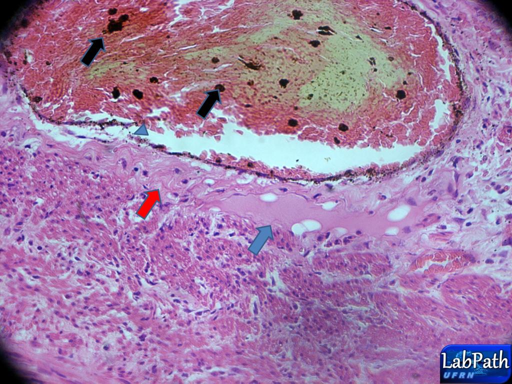
Notar na imagem um vaso
congesto(hiperemiado) afrouxa
devido a quantidade de hemácias e plasma(cabeça de seta azul) liberando
fibrina(seta vermelha) e plasma(seta azul).As setas pretas indicam a presença
de hemossiderina.
Translation: Notice in the image a vase congested (hyperemic) loosing because the amount of red blood cells and plasma (blue arrowhead) releasing fibrin (red arrow) and plasma (blue arrow). The black arrows indicate the presence of hemosiderin.
Translation: Notice in the image a vase congested (hyperemic) loosing because the amount of red blood cells and plasma (blue arrowhead) releasing fibrin (red arrow) and plasma (blue arrow). The black arrows indicate the presence of hemosiderin.

4x
Visualização detalhada(Details)
Notar que o infiltrado inflamatório já atingiu as
camadas musculares.
Translation: Note that the inflammatory infiltration has reached the muscle layers.
Translation: Note that the inflammatory infiltration has reached the muscle layers.

40x
Visualização detalhada(Details)
Notar a presença de PMNS,(cabeça de setas);as estrela mostra exsudato plásmatico.
Translation: Note the presence of PMNs (arrow head); star shows the plasma exudate.
Translation: Note the presence of PMNs (arrow head); star shows the plasma exudate.


Adorei esse post, muito ilustrativo. Parabéns
ResponderExcluir